Gel electrophoresis Gel electrophoresis. In this lesson you will learn how to use a DNA ladder to interpret experimental results.
 How Can Gel Electrophoresis Be Used To Detect Mutations Socratic
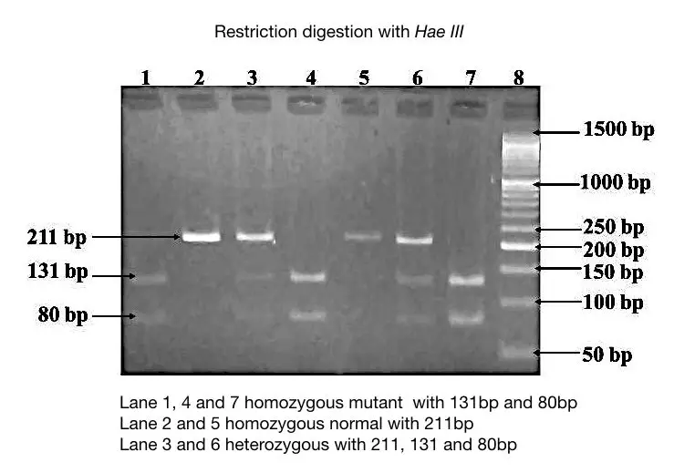
The location of the bands on a gel reveals the size of the DNA fragment.
How to read gel electrophoresis. The interpretation part of the agarose gel electrophoresis quite difficult if someone is a novice. The reaction is a free radical polymerization usually carried out. Electrophoresis through agarose or polyacrylamide gels is a standard method used to separate identify and purify biopolymers since both these gels are porous in nature.
The volume of the buffer should not be greater than 13 of the electrophoresis chamber. If doing a gel extraction in an 8-well gel combine 30 μL DNA. Prepare the samples by adding 6X loading buffer to each.
Read the previous part of this article. The UV light reveals the gel electrophoresis band intensity of the DNA or other molecular samples. Gel electrophoresis is used to analyze DNA restriction digest and ligation experiments.
Wear safety goggles and an apron. To identify the basic components of an electrophoresis system and to obtain a basic understanding of their functions. Print the picture of the gel on paper and get a ruler and a pencil.
After the gel solidifies the gel is submerged in a buffer-filled electrophoresis chamber which contains a positive electrode anode at one end and a negative electrode cathode at the other. Gel electrophoresis separates DNA fragments on the basis of size. Im amplyfing a DNA sequence using thermal cycler PCR.
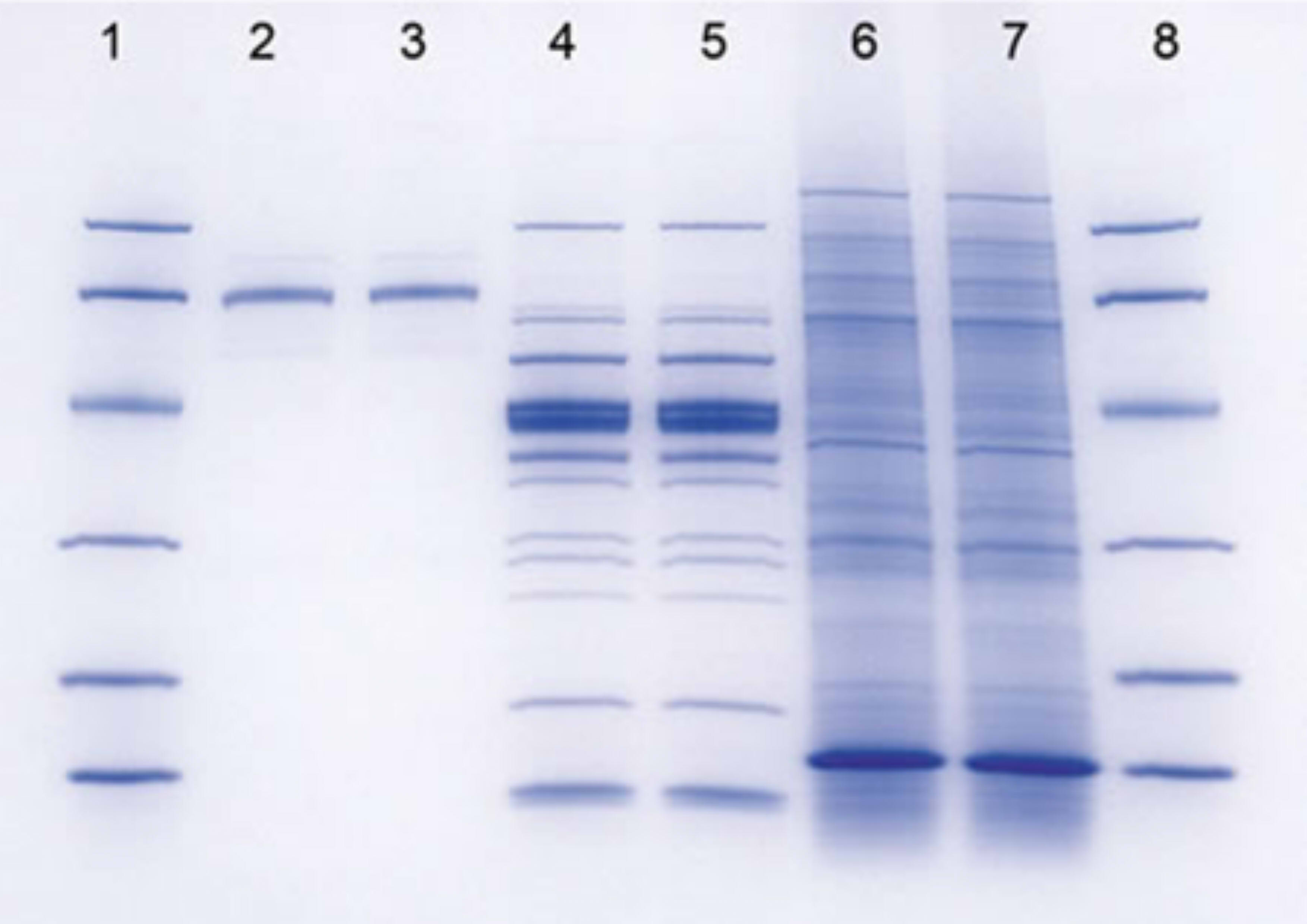
Gel electrophoresis is a technique used to separate DNA fragments or other macromolecules such as. Today well be talking about gel electrophoresis what is gel electrophoresis you might ask well its a lab technique usually used in the biochemistry lab for separating out DNA or proteins based on their size and lets talk about how it works so first you need to have the gel this can be made out of different kinds of substances such as agarose and polyacrylamide both of which Ill discuss. A complete guide for analyzing and interpreting gel electrophoresis results.
How Does It Work. The gel electrophoresis band intensity reveals the concentration of the molecule. Identify the size of each standard band in bp.
Look at the lane that contains the standard for the gel. Large ones cant get through the gel easily so they stay close to the top. DNA bands can only be visualized using agarose gel electrophoresis.
Enter that data into a column in Excel. Polyacrylamide gels are chemically cross-linked gels formed by the polymerization of acrylamide with a cross-linking agent usually NN-methylenebisacrylamide. Agarose gel electrophoresis is an important technique in molecular genetics for a long.
Visualizing the DNA fragments. NEVER PUT THE POWER SOURCE OR ELECTROPHORESIS CHAMBER NEAR RUNNING OR STANDING WATER. Read 3 answers by scientists to the question asked by Zaki Yamani on Oct 23 2017.
Ensure the gel is in the correct orientation with the negativeblack electrode above the wells so that the DNA runs toward the positivered electrode. When running the PCR product using gel electrophoresis I. In genomic research analyzing and interpreting the agarose gel electrophoresis results are very crucial.
Gel electrophoresis apparatus an agarose gel is placed in this buffer-filled box and an electrical field is applied via the power supply to the rear. For a 16-well gel combine 5 μL of DNA with 1 μL of 6X loading buffer in order to load 5 μL.


